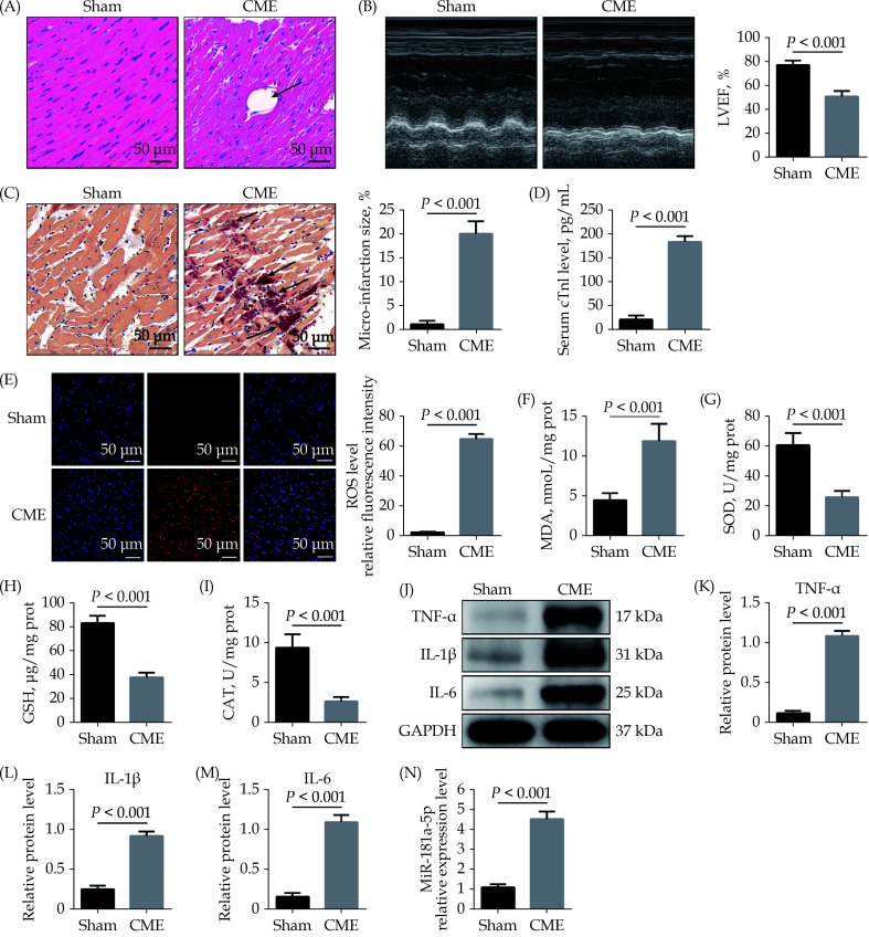
Figure 1.
Upregulation of miR-181a-5p in heart tissue of rats.
(A): Myocardial tissue samples stained with hematoxylin and eosin. Microspheres are marked using black arrows (× 200 magnification, scale bar = 50 µm); (B): echocardiography was used to assess cardiac function and to quantify LVEF (n = 10); (C): haematoxylin-basic fuchsin-picric acid staining was used to measure micro-infarction size (× 200 magnification, scale bar = 50 µm) (n = 6); (D): serum cTnI concentration in each group (n = 10); (E): the ROS production was detected by the dichloro-dihydro-fluorescein diacetate assay (× 200 magnification, scale bar = 50 µm) (n = 6); (F–I): myocardial MDA, SOD, GSH, and CAT level in each group (n = 6); (J–M): western blotting was used to quantify myocardial TNF-α, IL-1β, and IL-6 protein level, with glyceraldehyde 3-phosphate dehydrogenase as a loading control (n = 3); and (N): miR-181a-5p level was significantly elevated in heart tissue following CME (n = 10). Data were shown as mean ± SE (standard error) based on at least three independent experiments. CAT: catalase enzymes; CME: coronary microembolization; cTnI: cardiac troponin I; GSH: glutathione; IL-1β: interleukin-1β; IL-6: interleukin 6; LVEF: left ventricular ejection fraction; MDA: malondialdehyde; ROS: reactive oxygen species; SOD: superoxide dismutase; TNF-α: tumor necrosis factor-α.

