
Figure 4.
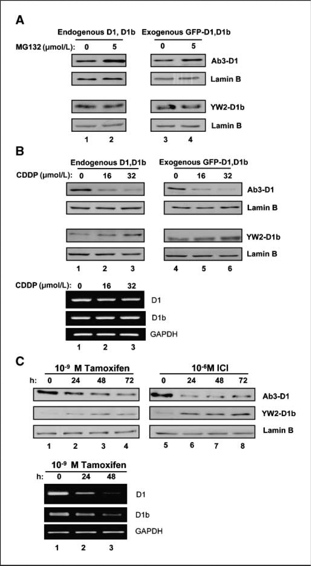
Distinct behaviors of cylin D1b protein compared with cyclin D1. A, MCF-7 cells were transfected with GFP–cyclin D1b or D1 expression plasmids. Cells were treated with 5 μm MG-132 for 6 h. Total cell lysate was resolved by SDS-PAGE. Endogenous D1 (left, top) and D1b (left, low) proteins were detected by immunoblotting. Exogenous GFP–cyclin D1 (right, top) and GFP–cyclin D1b (right, low) were blotted with GFP antibody. Lamin B was used as equal loading control. B, total protein or RNA was isolated from MCF-7 cells treated with CDDP for 16 h and resolved by SDS-PAGE. Western blot analysis for endogenous D1 (left, top), D1b (left, bottom), and exogenous GFP-D1 (right, top), GFP-D1b (right, bottom) was performed. Lamin B was used as a control for equal loading. RNA levels were evaluated by transcript specific RT-PCR, glyceraldehyde-3-phosphate dehydrogenase (GAPDH) is a control for equal loading (bottom). C, MCF-7 cells were treated with either 10−9 mol/L Tam or 10−6 mol/L ICI for up to 72 h. Total cell lysates were harvested at indicated intervals. The effects of tamoxifen (left) and ICI (right) upon expression of cyclin D1 and cyclin D1b protein were detected by Western blot. Lamin B was used as equal loading control. RNA levels were evaluated by transcript specific RT-PCR, GAPDH is a control for equal loading (bottom).