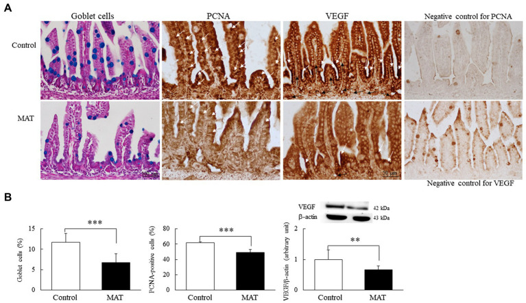
Figure 4.
Effects of MAT on (A) immunohistochemistry results and (B) results of quantitative analysis of goblet cells and PCNA-positive cells and Western blotting of VEGF in 7-day-old mice. The immunoreactivity for PCNA (white arrow) was localized to the nucleus and appeared along the epithelium of the intestinal mucosa and in the basal area of the intestinal crypts. Dark brown VEGF-positive staining (white arrow) was localized at the apical cytoplasm of the epithelial cells in both groups, and the VEGF-positive endothelial cells (black arrow) of blood vessels were observed in the lamina propria of the control group. The mice born to the control dams demonstrated more prominent immunoreactivity for PCNA and VEGF staining. The mice born to the antibiotic-treated dams exhibited significantly fewer mucus-positive goblet cells (blue stained) and PCNA-positive cells and significantly lower VEGF protein expression than did the mice born to the control dams (n = 9). **p < 0.01; ***p < 0.001. MAT, maternal antibiotic treatment; PCNA, proliferating cell nuclear antigen; and VEGF, vascular endothelial growth factor.

