Fig. 3.
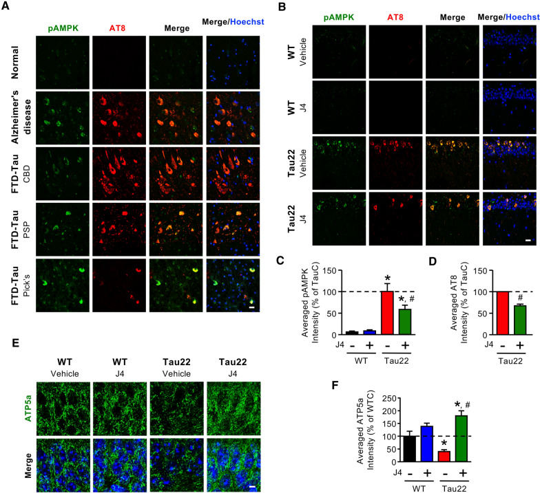
Chronic J4 treatment decreases AMPK activation and rescues mitochondrial abnormalities in the hippocampi of Tau22 mice. a Posterior hippocampal sections (6 μm) from normal subjects and Alzheimer’s Disease and FTD-Tau (CBD, PSP, and Pick’s disease) patients were subjected to IHC staining. The levels of phospho-AMPK and hyperphosphorylated tau were evaluated by staining with the indicated antibodies (pAMPKThr172, green; AT8 for pTauSer202/Thr205, red). b–f Mice were treated as indicated (WTC, black; WTJ, blue; TauC, red; TauJ, green; n = 5–7 in each group) from the age of 3–11 months. Hippocampal sections (20 μm) were prepared and subjected to IHC staining using the indicated antibodies (pAMPKThr172, green; AT8 for pTauSer202/Thr205, red), and the staining was quantified (c, d). Scale bar, 20 μm. e, f The level of the mitochondrial marker ATP5a was evaluated by staining with an anti-ATP5a antibody (e, green) and quantified (f; n = 3 in each group). Scale bar, 5 μm. The data are expressed as the mean ± S.E.M. *p < 0.05 versus the WTC group; #p < 0.05, versus the TauC group, one-way ANOVA

