. 2021 May 5;12(24):8333–8341. doi: 10.1039/d1sc00522g

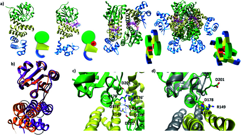
Fig. 1. (a) TtCarH structures and their cartoon representations, colored by their domain: B12-binding (green), 4-helix bundle (yellow), DNA-binding (blue), and AdoCbl (magenta). L–R: apo-monomer (representative from MD simulations), holo-monomer, holo-dimer, and holo-tetramer (all PDB: 5C8D). (b) 4-helix bundle (bottom) positions in each monomer aligned to the B12-binding domain (top): holo-TtCarH (orange, PDB: 5C8D) and apo-TtCarH (purple, simulated). A different color scheme to panel (a) is used to distinguish between different structures. Arrows indicate helical displacement in apo-TtCarH relative to holo-TtCarH (see also Fig. S3a and b). (c) Close-up of the interface between monomer units in the holo-TtCarH head-to-tail dimers with salt-bridges identified here (D178–R149) and previously10 (D201–R176) highlighted. (d) The same interfacial region illustrated in panel (c), but now of the simulated apo-TtCarH monomer. The change in 4-helix bundle conformation relative to holo-TtCarH (in grey) results in a helix moving into the dimer interface such that the key salt-bridge residues are no longer able to stabilize the head-to-tail dimers (see also Fig. S3c).

Fig. 1