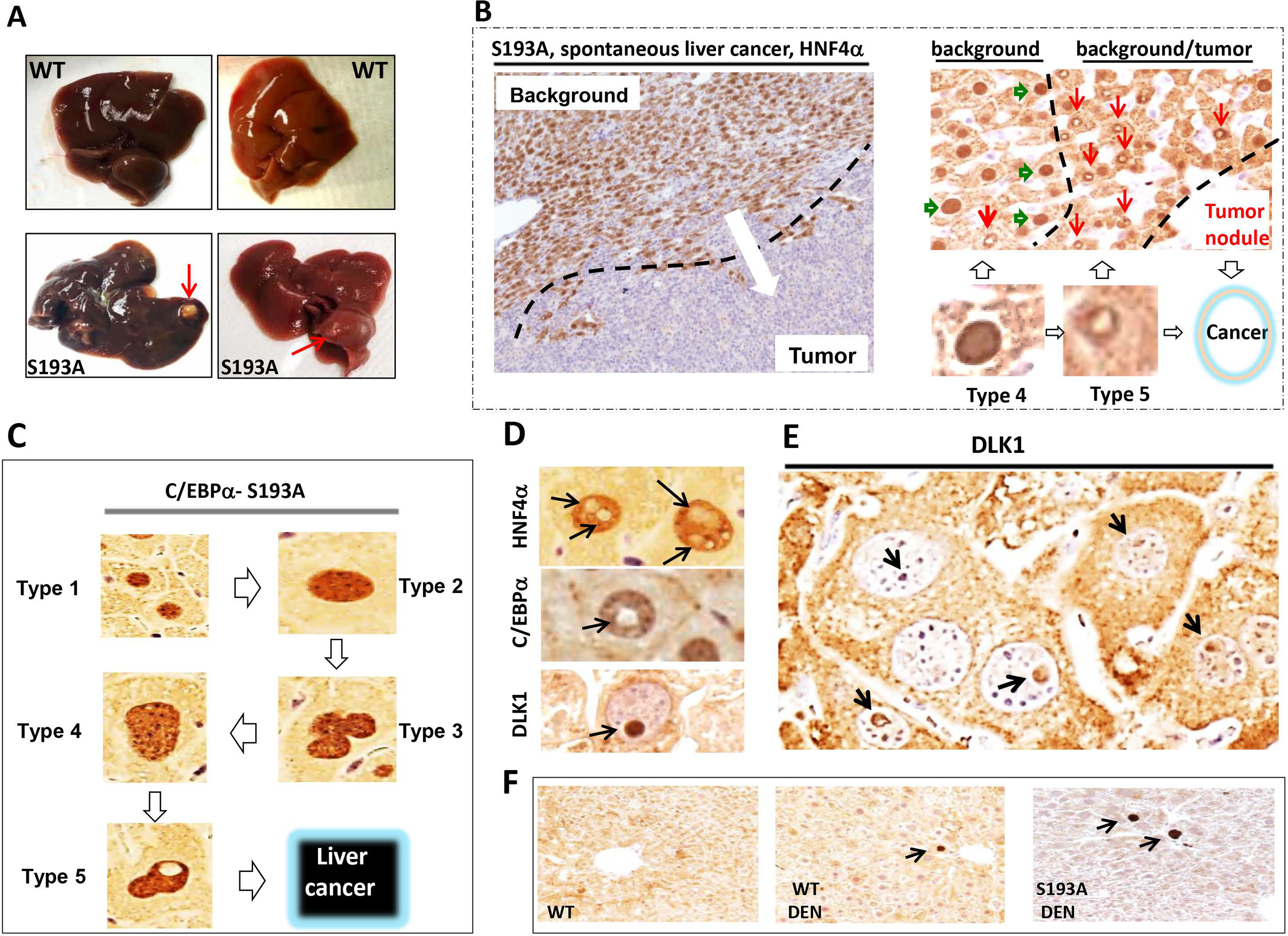
Figure 5. C/EBPα-S193A mice develop spontaneous liver cancer, background of which is characterized by abundant type 4 and 5 hepatocytes.

(A) Typical picture of liver cancer in 17 month-old S193A mice. Red arrows show large external tumor nodules. (B) Typical picture of HNF4α staining of the liver tumor in S193A mice. Background section is positive for HNF4α; tumor section is negative. Right image shows HNF4α staining of a border region between tumor section and background of livers of S193A mice. Hepatocytes type 4 (green arrows) are observed far from the tumor section, while hepatocytes type 5 (red arrows) are in very close proximity to the area with cancer. (C) A summary of studies of hepatocytes within tumor nodules of DEN-treated S193A mice and within tumor nodules of spontaneous liver cancer in S193A mice. (D) Staining of S193A hepatocytes of DEN-treated mice (28 wks) with markers of hepatocytes HNF4α and C/EBPα and with Stem Cell Marker DLK1. (E) A bigger field of the liver with hepatocytes that have DLK1-positive intra-nuclear inclusions. (F) Background sections of DEN-treated S193A mice were stained with Abs to DLK1.
