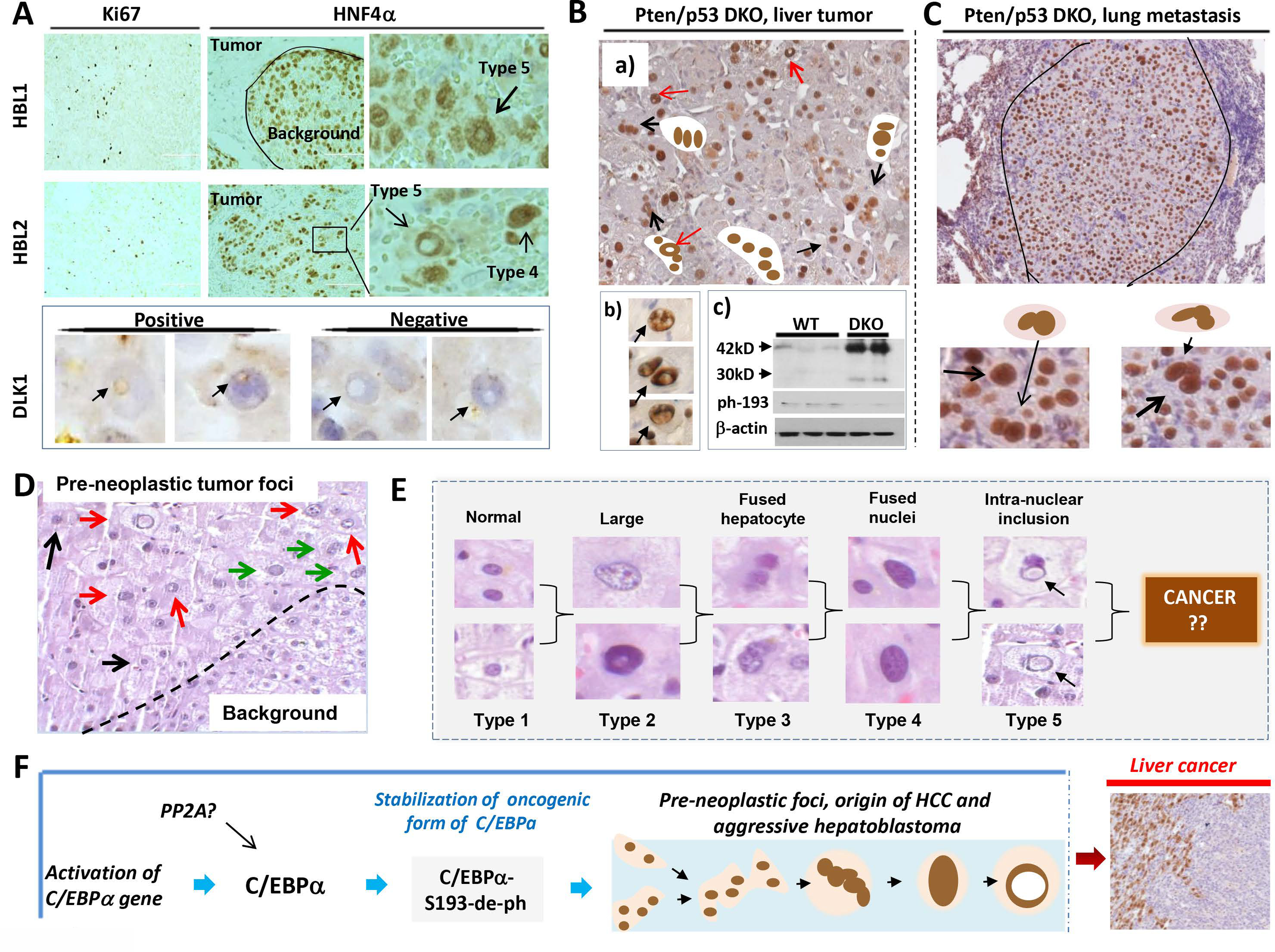
Figure 8. Tumor initiating hepatocytes are abundant in patients with aggressive HBL, in liver tumor sections of Pten/p53 DKO mice and in a patient with high risk for development of liver cancer.

(A) ki67 and HNF4α staining of livers from patients with aggressive hepatoblastoma. Types 4 and 5 hepatocytes are shown by arrows on the enlarged images (right). Bottom panel shows staining with stem cell marker DLK1. Positive and negative type 5 hepatocytes are shown. (B) a): Typical picture of HNF4α staining of tumor nodules of Pten/p53 DKO mice. Multi-nucleated and type 5 hepatocytes are shown. Section b) shows enlarged images of PTIHs in the tumor nodules of Pten/p53 DKO mice. Section c) shows Western blotting with antibodies to total C/EBPα and to ph-S193-C/EBPα. (C) HNF4α staining of a lung metastasis in Pten/p53 DKO mice. Bottom shows enlarged images of PTIHs in the lung metastasis. (D) H&E staining of the liver of a patient who has high risk for development of liver cancer. Pre-neoplastic tumor foci and background areas are shown. This section of pre-neoplastic foci contains type 2 (black arrows), type 4 (green arrows) and type 5 (red arrows) hepatocytes. (E) Summary for all types of hepatocytes observed in pre-neoplastic foci of a patient with high risk of liver cancer. (F) The main hypothesis for the origin of aggressive HBL and HCC (see text).
