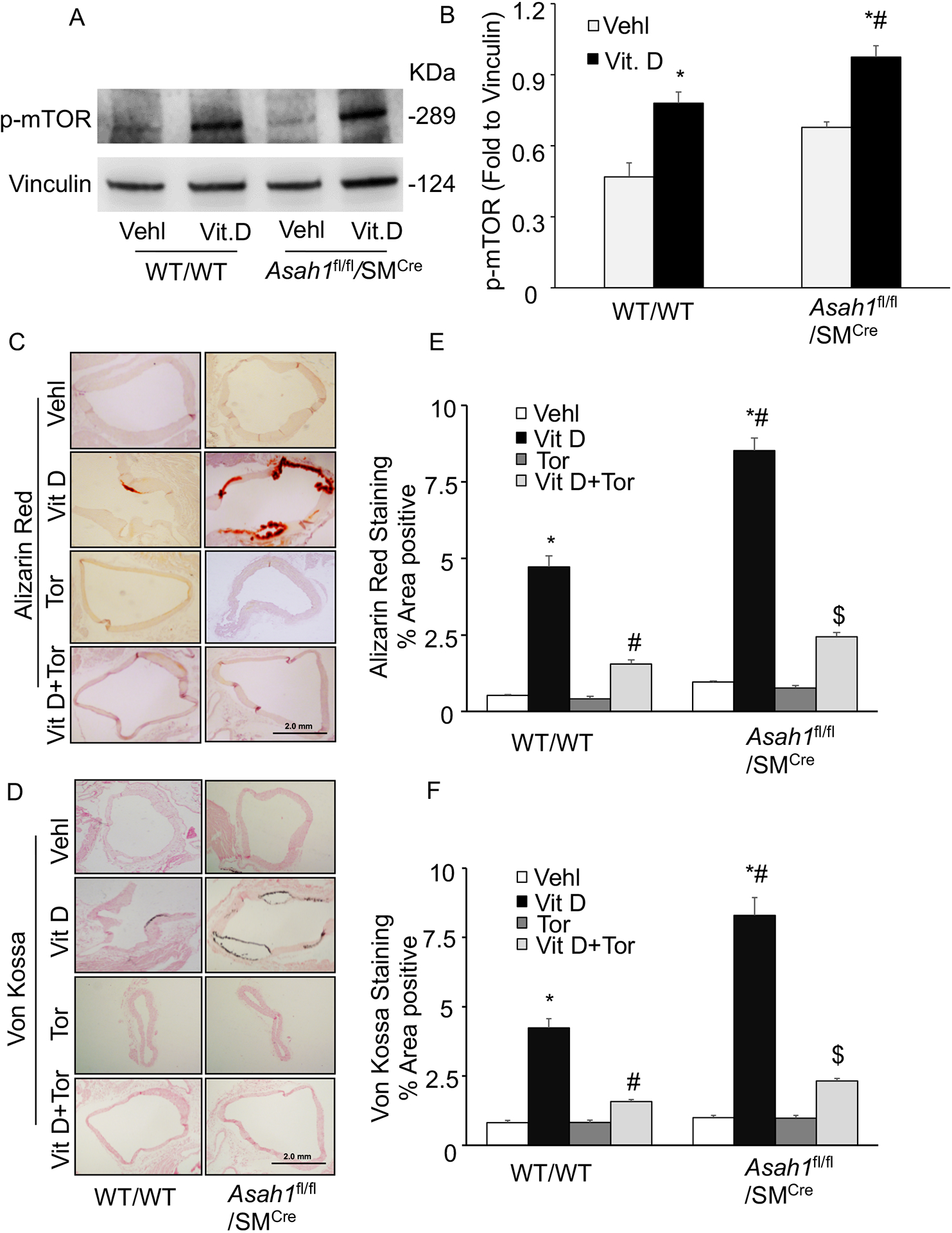
Figure 2.

Effect of mTOR-inhibition on AMC in the Asah1fl/fl/SMCre mice. (A, B) Western blot analysis of enhanced expression of p-mTOR (Ser2448) in Vit D-treated Asah1fl/fl/SMCre mice. n=4. Representative photomicrographs showed (C) calcium deposition by Alizarin Red S staining (red color) and (D) mineral deposition by Von Kossa staining (black color). (E, F) Summarized bar graph showed Torin-1 significantly decreased calcium deposition and mineralization in aortic medial wall. n=5, SMC: smooth muscle cell; Vehl: vehicle; Vit D: vitamin D; Tor: Torin-1. ‘n’ is mouse number. *P < 0.05 vs WT/WT Vehl; #P < 0.05 vs WT/WT Vit D group; $P< 0.05 vs. Asah1fl/fl/SMCre Vit D group by one-way ANOVA with Holm-Sidak’s test post-hoc analysis. Data are shown as mean ± SEM of values.
