. 2021 May 18;12(24):8288–8310. doi: 10.1039/d1sc01359a

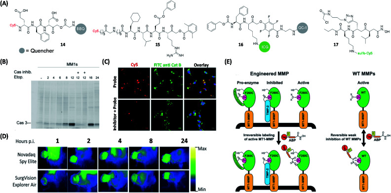
Fig. 6. Protease qABPs for visual labeling of different proteases in live cells and mouse tumors. (A) Structures of reported ABPs to study proteases. (B) In-gel fluorescence analysis of Casp3 in MM1s cells treated with 14. The addition of Casp3 inhibitor blocked the labeling of Casp3 by the probe, which demonstrated the specificity of the probe to Casp3. Reproduced from ref. 64 with permission from Royal Society of Chemistry, copyright 2016. (C) Labeling of cathepsin B in non-small lung cancer cells from patient using 15 and anti-cathepsin B FITC antibody. The labeling of cathepsin B using 15 was blocked by the addition of cathepsin B inhibitor, but the inhibitor did not influence the labeling efficiency of the antibody. Reproduced from ref. 66 with permission from Royal Society of Chemistry, copyright 2019. (D) Still images from Novadaq Spy Elite and SurgVision Explorer Air of 4T1 tumors at different time points after injection of 16, which showed the potential of 16. Reproduced from ref. 69 with permission from Springer, copyright 2020. (E) Specific labeling of active MMP-14 using protein engineering coupled with 17. The ABP only labeled active MMPs and cannot label enzymes that are inaccessible to zinc or their inhibitory forms. Reproduced from ref. 75 with permission from American Chemical Society, copyright 2018.

Fig. 6