. 2021 May 18;12(24):8288–8310. doi: 10.1039/d1sc01359a

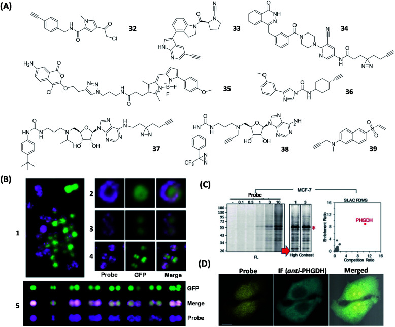
Fig. 9. ABPs and AfBPs designed based on specific enzyme inhibitors and their labeling of enzymes in cells. (A) Structures of reported enzyme inhibitor-based ABPs. (B) Confocal images of S. aureus Newman labeled with 35 (1). Confocal images of S. aureus Newman-GFP cell labeled with 35 during exponential phase (2 and 3) and stationary phase (4 and 5). 35 successfully demonstrated that FphB is concentrated in the isolated membrane of bacterial cells. Reproduced from ref. 109 with permission from Springer Nature, copyright 2018. (C) Proteome profiles of MCF-7 cells labeled with 39, with a visible FL band around 55 kDa. These demonstrated the specific labeling of PHGDH by 39 in living cells. (D) Confocal images of MCF-7 cells treated with 39 and anti-PHGDH. Reproduced from ref. 115 with permission from Wiley-VCH GmbH, copyright 2018.

Fig. 9