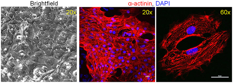
Figure 5. iPSC derived cardiomyocytes at Day 16 of differentiation.
More than 90% of the cells show positive staining for the cardiomyocyte marker α-actinin. iPSC derived cardiomyocytes exhibit sarcomere structure by immunofluorescent microscopy at high magnification (60x). Scale bars, 100 μm (brightfield); 20 μm.

