. 2021 Jun 8;9(24):4906–4914. doi: 10.1039/d0tb02762f

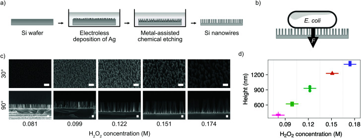
Fig. 1. Fabrication of silicon nanowires for bacterial interfacing. (a) Illustration of silicon nanowire fabrication process. (b) Illustration of nanowire interfacing mechanism with E. coli, where the force F is either the result of cells settling under gravity or increased by centrifugation. (c) SEM micrographs showing the impact of varying the concentration of hydrogen peroxide (H2O2) in the etchant during MACE process. Samples imaged at 30° or 90°, as indicated, with no tilt correction applied. Scale bars are 1 μm for 30° and 200 nm for 90° images. (d) Impact of hydrogen peroxide concentration on resulting nanowire height. Horizontal lines represent the median of measurements. n = 5.

Fig. 1