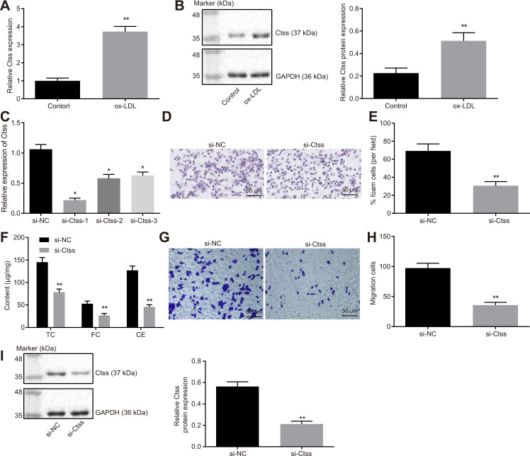
Figure 6.
Ctss affects BMDM expression of atherosclerotic phenotype. (A) The mRNA expression of Ctss in BMDMs after 24-h treatment with ox-LDL determined by RT-qPCR. (B) The protein expression of Ctss in BMDMs after 24-h treatment with ox-LDL determined by Western blot analysis. (C) Silencing efficiency of Ctss after 24-h treatment with ox-LDL and different siRNA detected by RT-qPCR. (D–E) The proportion of foam cells in BMDMs treated with si-Ctss by oil red O staining (200 ×) (Five visual fields were randomly read and photographed, and the mean value was obtained. Each experiment was repeated three times). (F) Serum TC, FC and CE levels in BMDMs treated with si-Ctss determined by ELISA. (G, H) BMDM migration in response to the treatment of si-Ctss evaluated by Transwell assay (200 ×) (Five visual fields were randomly read and photographed, and the mean value was obtained. Each experiment was repeated three times). (I) Ctss protein expression in BMDMs in response to the treatment of si-Ctss evaluated by western blot analysis. ** p < 0.01 vs. the BMDMs treated with NC mimic, si-NC or without treatment. Statistical data were measurement data, and described as mean ± standard deviation. The paired t test was used for comparisons between two groups. The one-way analysis of variance was adopted for comparisons among multiple groups, followed by Tukey’s post hoc test. The experiment was repeated 3 times independently.

