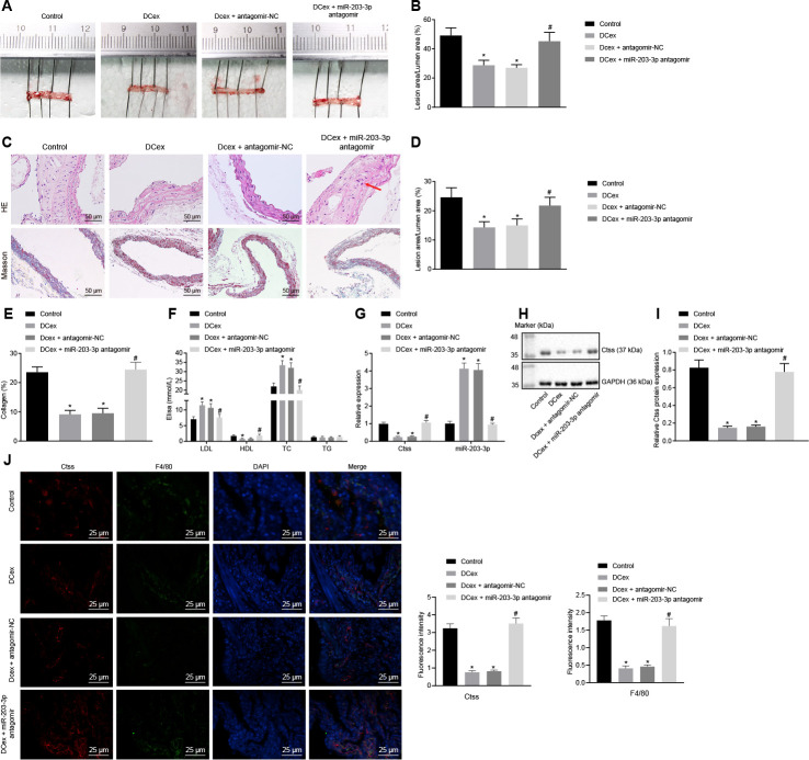
Figure 8.
DCex and miR-203-3p contribute to alleviate AS in vivo. The HFD-fed ApoE-/- mice did not receive any treatment as controls, or were treated with DCex alone, DCex + antagomir-NC, or miR-203-3p antagomir (n = 8 for each group). (A, B) Oil red O staining for atherosclerotic plaque in ApoE-/- mice. (C–E) HE staining (200 ×) and Masson staining (200 ×) for atherosclerotic plaque in ApoE-/- mice (In panel C, red arrows show plaque location). (F) Serum LDL, HDL, TC, and TG levels in ApoE-/- mice determined by ELISA. (G) miR-203-3p expression and mRNA expression of Ctss in vascular tissues in ApoE-/- mice evaluated by RT-qPCR. (H, I) The protein expression of Ctss in vascular tissues in ApoE-/- mice evaluated by Western blot analysis. (J) Immunofluorescent double staining for the expression of F4/80 and Ctss (400 ×). * p < 0.05 vs. mice without treatment; # p < 0.05 vs. mice treated with DCex. Statistical data were measurement data, and described as mean ± standard deviation. The one-way analysis of variance was adopted for comparisons among multiple groups, followed by Tukey’s post hoc test. The experiment was repeated 3 times independently.

