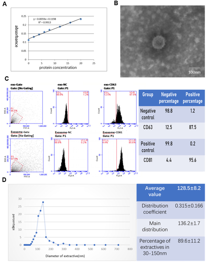
Figure 2.
Identification of exosomes deprived from hAECs. (A) Protein concentration curve from the BCA test. (B) The morphology of exosomes was observed under an electron microscope. (C) The phenotype of exosomes for CD63 and CD81 was identified by flow cytometry. (D) The diameter distribution of exosomes was measured by nanoparticle tracking analysis.

