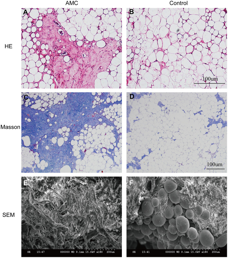
Figure 2.
Histological structure analysis of AMC and Coleman fat. (A, B) Hematoxylin/eosin staining of AMC and Coleman fat (control) before grafting. (C, D) Masson’s trichrome staining of AMC and Coleman fat (control) before grafting. (E, F) Microstructure of AMC and Coleman fat (control) before grafting, as determined by electron microscopy.

