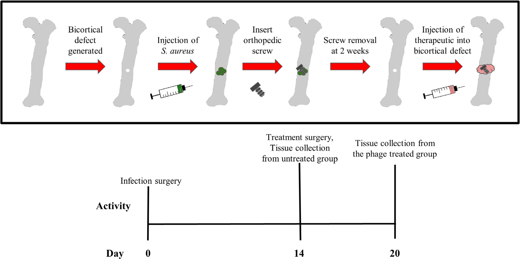
FIGURE 2.

Overview of in vivo osteomyelitis model. Day 1: Staphylococcus aureus (S. aureus) was injected into the bone defect and screw was placed. Day 14: contaminated screw was removed, bacteriophage treatment and a new (sterile) screw were applied to the treatment group; tissues from the untreated group were collected for bacterial counting. Day 20: treatment group was imaged, and tissues were collected for bacterial counting
