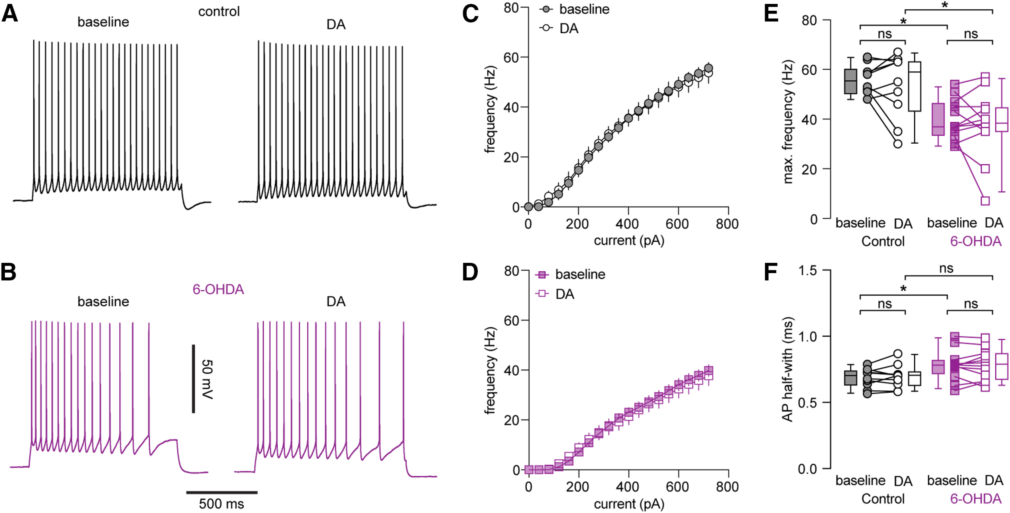
Figure 8.

DA application could not rescue the decreased excitability of PTNs following loss of midbrain DA neurons. A, B, Representative AP traces of PTNs from controls and 6-OHDA mice in the absence and presence of DA. C, D, Frequency-current curves of PTNs from controls (C) and 6-OHDA mice (D) in the absence and presence of DA. E, Summarized graphs showing the lack of impact of DA on maximal firing frequency of PTNs from controls and 6-OHDA mice. F, Summarized graphs showing the lack of impact of DA on AP width of PTNs from controls and 6-OHDA mice; ns, not significant; *p < 0.05, WSR or MWU tests.
