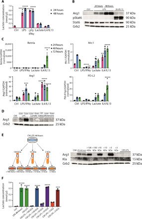
Fig. 1. Inflammation-associated lactate and M2-like gene expression are biochemically uncoupled.

(A) Intracellular lactate concentration was determined in wild-type BMDMs left untreated (Ctrl), and LPS/IFNγ-, LPS-, lactate-, or IL4/IL13-treated for 24 or 48 hours. Data shown are representative of three independent experiments. (B) BMDMs, which were treated with solvent (Ctrl), LPS, or IL4/IL13, were used to isolate whole-cell lysates and analyzed by Western blotting for the indicated proteins. (C) RNA from wild-type BMDMs left untreated (Ctrl), stimulated with LPS/IFNγ, lactate, or IL4/IL13 was used for quantitative reverse transcription polymerase chain reaction. Data shown are the mean fold increase of the 24-hour Ctrl group. GAPDH, glyceraldehyde-3-phosphate dehydrogenase. (D) Wild-type BMDMs were stimulated with LPS, lactate, or rotenone. DMSO, dimethyl sulfoxide. (E) Wild-type BMDMs were stimulated with LPS for 48 hours (LPS 48 hours). Supernatant was used unfractionated (CM LPS) or fractionated for 100-, 10-, or 3-kDa protein size. Unfractionated and fractionated supernatant was transferred to naïve BMDMs for further 24 hours. (F) Intracellular lactate concentration was determined in the described groups. Statistically significant differences were determined by one-way or two-way (A and C) analysis of variance (ANOVA) with Tukey correction; n = 2 biological replicates. All values are means ± SEM; *P < 0.05; **P < 0.01; ***P < 0.001; ****P < 0.0001. Superscripts indicate statistical significance compared to the control group. If not indicated otherwise, n = 3 biological replicates.
