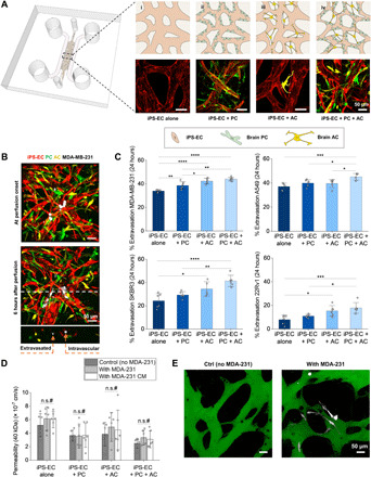
Fig. 1. The presence of PCs and ACs in the MVNs promotes TC extravasation but decreases barrier permeability.

(A) Schematic of the microfluidic chip used in the study to generate four MVN platforms from the sequential addition of PCs and ACs to iPS-ECs. (B) MDA-MB-231 (MDA-231) cells perfused in the triculture MVNs and left to extravasate for 6 hours. Examples of extravasated and intravascular TCs are shown. (C) Extravasation efficiencies of breast MDA-231, breast SKBR3, lung A549, and prostate 22Rv1 in the four MVN platforms at 24 hours after perfusion. (D) MVN permeability to 40 kDa of dextran in the four platforms considered. Permeability is also assessed in the presence of MDA-231 TCs and their conditioned media (CM). (E) Representative images of triculture MVNs perfused with 40-kDa fluorescein isothiocyanate–dextran (green) with and without MDA-231 TCs. *P < 0.05, **P < 0.01, ***P < 0.001, and ****P < 0.0001; not significant; n.s.#, not significant across all conditions (pairwise).
