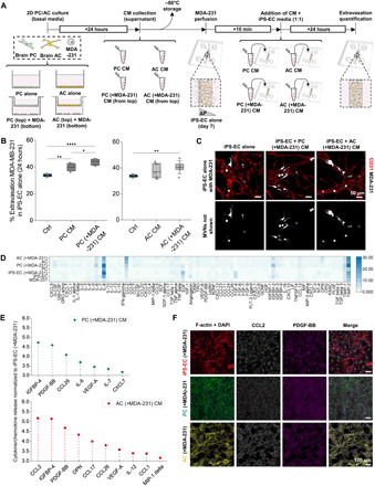
Fig. 2. PC- and AC-secreted factors increase the extravasation potential of TCs.

(A) Schematic of the collection of PC and AC CM and subsequent treatment of the MVNs before TC perfusion. (B) Extravasation efficiencies of breast MDA-231 in monoculture MVNs with or without CM treatment at 24 hours after perfusion. (C) Representative images of breast MDA-231 in monoculture MVNs with or without CM treatment at 24 hours after perfusion. (D) Heatmap for cytokine array showing relative magnitudes of secreted factors from MDA-231, iPS-EC, PC, and AC alone, as well as iPS-EC, PC, and AC cultured in Transwell inserts with MDA-231 cells after 24 hours of culture. (E) Relative magnitude of significantly up-regulated cytokines/chemokines by PC or AC cultured in inserts with MDA-231 compared to iPS-EC cultured with MDA-231.(F) Representative images of 2D immunofluorescence staining for F-actin + DAPI, CCL2, and PDGF-BB in iPS-ECs, PCs, and ACs, cultured with MDA-231 in Transwell inserts. *P < 0.05, **P < 0.01, ***P < 0.001, and ****P < 0.0001; not significant; n.s.#, not significant across all conditions (pairwise).
