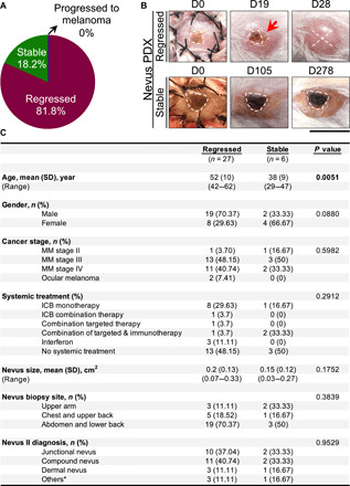
Fig. 1. Most of the melanocytic nevus PDXs undergo regression, which is associated with increased participant age.

(A) Distribution of nevus PDX outcomes (regressed, stable, or progressed to melanoma, n = 33 mice). (B) Representative images of regressed nevus PDX at days 0, 19, and 28 and stable nevus PDX at days 0, 105, and 278 after transplant. Dashed lines outline the melanocytic nevus on the human skin graft, “X” signifies nevus rejection, and the red arrow points to the inflammation in the regressing nevus; scale bar, 1 cm. (C) Participant demographic information associated with regressed and stable nevus PDXs (n = 33). Mann-Whitney U test is used for age and size variables, and Pearson’s χ2 tests are used for other categorical variables. *Others, benign melanocytic proliferations; MM, malignant melanoma; ICB, immune checkpoint blockade. Photo credit: Erik B. Schiferle, Massachusetts General Hospital.
