
Fig. 2. Melanocytic nevus PDX regression is associated with a massive expansion of human CD4+ T cells in the regressed nevi, the apoptosis of nevus melanocytes, and expansion of nevus-derived human T cells in the lymph node and spleen of the transplanted mice.
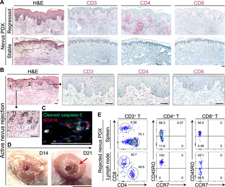
(A) Representative images of hematoxylin and eosin (H&E), human CD3, CD4, and CD8 stained histological sections of regressed and stable nevus PDXs. Scale bar, 100 μm. (B) Representative images from H&E- and CD3/CD4/CD8-stained sections of actively rejecting nevus PDX. Insets show (1) the site of active nevus nest rejection and (2) immune cell infiltration. Scale bars, 100 μm. (C) Representative immunofluorescence (IF) image of cleaved caspase-3 (green) and SOX10 (red) in an actively rejecting nevus PDX at day 14 after transplant. Scale bar, 100 μm. (D) Representative macroscopic images of actively rejecting nevus PDX at days 14 and 21 after transplant. Dashed lines outline the area of nevus before rejection on the human skin, and arrow points to the inflammation and scabbing. Scale bar, 1 cm. (E) Representative flow cytometry plots of human CD4+ and CD8+ T cells in the spleen and lymph node of the rejected nevus PDX. CD45RO versus CCR7 expression shows the “effector memory” status of the rejected nevus-derived human T cells in the lymphoid organs of the NSG mice; numbers on the plots represent the percent cells within each gate. Photo credit: Erik B. Schiferle, Massachusetts General Hospital.