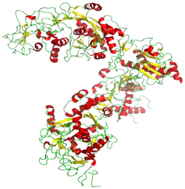
Figure 11.

Cartoon representation of SARS-CoV-2 nsp10-nsp16 complex structure showing loops (green), β-sheets (yellow), and helices (red) (PDB 6W4 H). Nsp10’s positively charged and hydrophobic surface interacts with a hydrophobic pocket and a negatively charged nsp16 surface, which helps to stabilize the SAM binding site.
