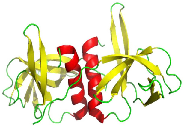
Figure 13.

Cartoon representation of crystal structure of SARS-CoV-2 nsp9 dimer structure showing loops (green), β-sheets (yellow), and helices (red) (PDB 6WXD). The inter-subunit interactions to form a dimer are due to van der Waals interactions between the interfacing copies of α1 helix C-terminal as a result of self-association of GxxxG protein-protein binding motif. 77
