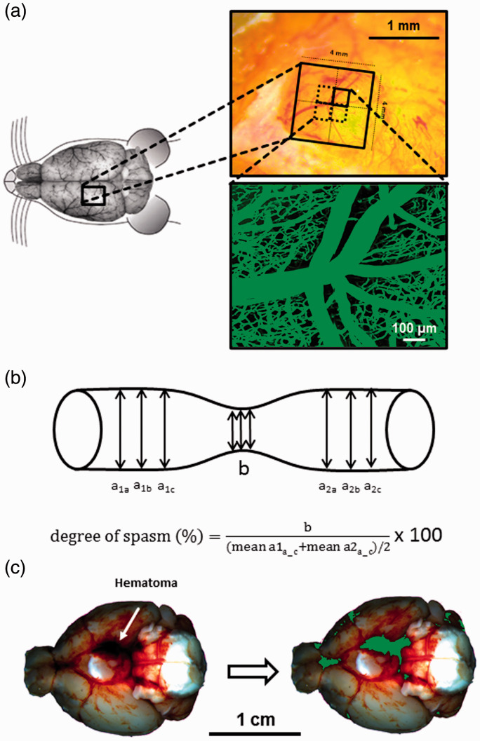
Figure 1.
Intravital microscopy - Experimental setup. (a) Schematic overview of cranial window placement for intravital microscopy. (b) Assessment of microvasospams formation and severity. (c) Example of a hematoma at the skull base after SAH (left) and the quantification of hematoma size (green) by an automated algorithm (right).

