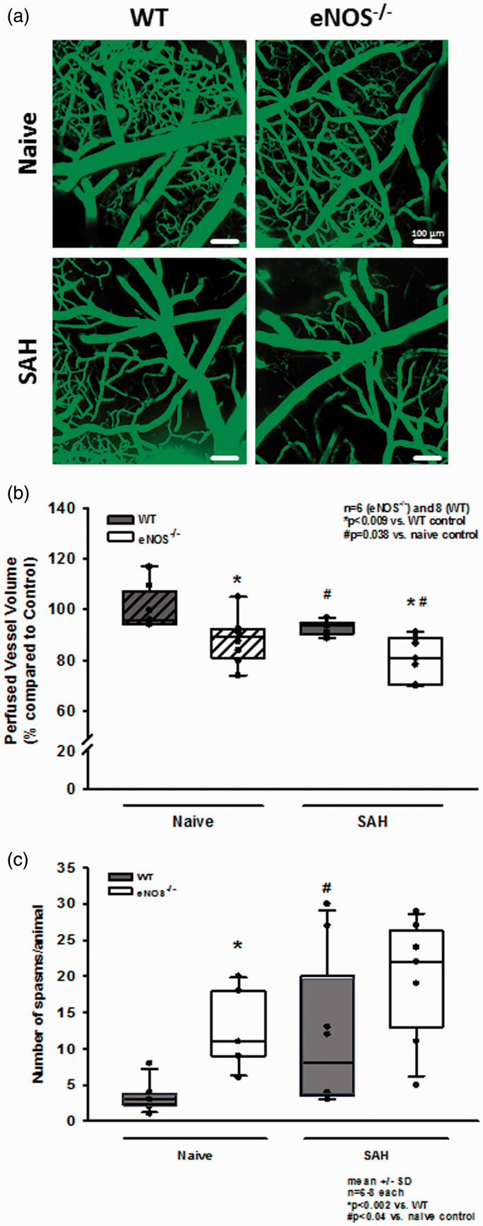
Figure 5.
Changes of the cerebral microcirculation after SAH. (a) Exemplary IVM screenshots depicting the cerebral microcirculation in naïve animals (upper panels) and after induction of SAH (lower panels). eNOS deficient mice already show a slight narrowing and rarefication on the capillary level. After SAH, there is (increased) microvascular constriction in both genotypes. (b) Corresponding to the exemplary picture, total perfused vessel volume is slightly but significantly lower in eNOS–/– homozygous animals compared to wild type mice. After SAH, perfused vessel volume is significantly lower in both genotypes, indicating relevant microvascular constriction and – thus – microcirculatory dysfunction. The drop in perfused vessel volume, however, is significantly more pronounced in eNOS–/– than in wt. (c) Microvasospasms occur very rarely under physiological conditions in wild type controls (left side, dark bar). The number of MVS in eNOS–/– is significantly elevated. After SAH, there is significant spasm formation in wt mice (right side of panel). This increase is more pronounced in eNOS–/– mice (striped bars).

