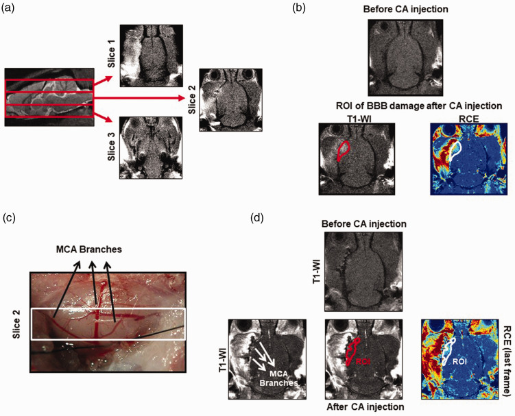
Figure 2.
ROI selection details to determine BBB damage and vascular parameters by DCE-MRI. (a) Images showing the 3 coronal sections with 3 mm of thickness in which the brain has been divided to analyze the vascular parameters and the BBB damage by the DCE-MRI technique. (b) T1-W and RCE images showing the ROI selected in the last frame of our DCE-MRI protocol, after CA injection to determine the BBB damage. (c) Image of the brain that correspond to slice 2 of our DCE-MRI analysis, where the transient occlusion of the MCA was performed. (d) T1-W and RCE images showing the ROI selected to determine by DCE-MRI the vascular density/function inside the ischemic brain.

