Figure 1.
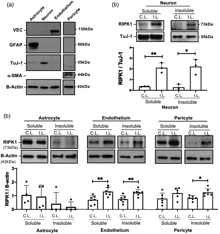
Cell-specific activation of RIPK1 after intracerebral hemorrhage (ICH). (a) Purity of isolated cell populations assessed by western blot using antibodies to glial fibrillary acidic protein (GFAP), neuron-specific class III beta-tubulin (TuJ-1), vascular endothelial cadherin (VEC), and alpha-smooth muscle actin (a-SMA). (b) RIPK1 was increased in both soluble and insoluble cell fractions in neurons isolated from ipsilateral (I.L.) hemispheres of wild-type (WT) mice 12 hours after ICH (**p < 0.005, *p < 0.05 vs. contralateral (C.L.) hemispheres, n = 3/group). (c) Increased RIPK1 in the detergent-insoluble fraction was detected in endothelium and pericytes isolated from ipsilateral hemispheres at 24 hours after ICH (**p < 0.005 vs. contralateral hemisphere, n = 7/group and *p < 0.05 vs. contralateral hemisphere, n = 6/group), but not in astrocytes (n = 3/group, p = ns, Mann Whitney U-test).

