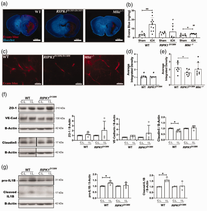
Figure 3.
RIPK1 and Mlkl contribute to blood-brain barrier damage after intracerebral hemorrhage (ICH). (a) Representative photomicrographs (1X) of the ICH lesion at 24 h showing Evans Blue extravasation (red). (b) Spectrophotometric quantification of extravasated Evans blue (EB) in ipsilateral hemispheres of wild-type (WT), RIPK1D138N/D138N, and Mlkl−/− mice. (**p < 0.005 and *p < 0.05 vs. sham; #p < 0.05 and ##p < 0.005 vs. WT ICH, n = 6 sham and 6-12 ICH/group). (c) Left panel: Representative photomicrographs of injured striatum at 30 d after ICH showing Evans blue dye (red fluorescence) leakage from blood vessels. Average difference in mean gray values of ipsilateral – contralateral color images from sham injured (d) and ICH (e) mice at 30 days. #p ≤ 0.05 vs. the corresponding ICH group; *p < 0.05 (n = 6-8/group). (f) Left panel: Representative Western blot showing changes in expression of proteins associated with blood-brain barrier integrity at 24 h after ICH. Right panel: Claudin 5 expression was significantly reduced in ipsilateral striatal brain homogenates of wild type (WT) mice compared to RIPK1D138N/D138N (*p < 0.05, n = 4/group). (g) Left panel: Representative western blot showing changes in interleukin-1 beta at 24 h after ICH in striatal brain homogenates. Right panel: Densitometry analyses of Western blot data showing increased pro- and cleaved forms of interleukin-1 beta in ipsilateral vs. contralateral striatum of wild type but not RIPK1D138N/D138N mice (*p< 0.05, n = 4/group).

