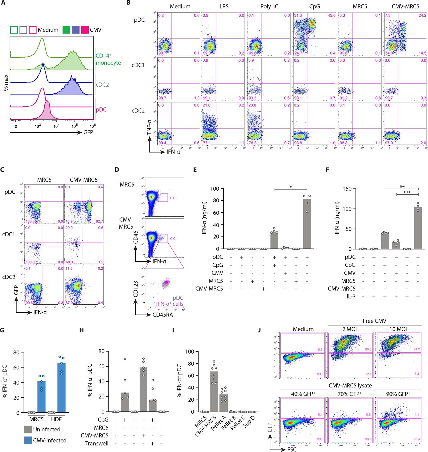
Fig. 1. pDCs produced interferon upon contact with live CMV-infected cells.

(A) The expression of GFP in purified human pDCs, cDC2 or CD14+ monocytes incubated with free CMV-GFP at 10 MOI for 36 hours.
(B) The expression of IFN-α and TNF-α in DC subsets stimulated with the indicated stimuli including MRC5 cells alone or infected with CMV (CMV-MRC5). Enriched DCs were cocultured with MRC5, and each plot was gated for pDC (CD123+ CD45RA+), cDC1 (CD11c+ CD141+) and cDC2 (CD11c+ CD1c+).
(C) The expression of IFN-α and GFP (as an indicator of CMV infection) in DC subsets cocultured with or without CMV-MRC5 and gated as in panel A. Representative of two experiments.
(D) The expression of IFN-α in total PBMC cocultured with or without CMV-MRC5. Right panel shows gated IFN-α+ cells (pink) overlaid onto gated CD123+ CD45RA+ total pDCs (grey).
(E-F) The concentration of IFN-α protein measured by ELISA in the supernatant of enriched pDCs stimulated with CpG, free CMV (10 MOI), MRC5 or CMV-MRC5 for 24 hours in the absence of IL-3 (E) or presence of IL-3 (F).
(G) Percentage of IFN-α+ pDCs cocultured with CMV-MRC5 or primary human dermal fibroblasts (HDF), infected at 0.5 MOI and cultured for 2 or 5 days, respectively.
(H) Percentage of IFN-α+ pDCs cocultured with CMV-MRC5 or CpG in transwell cultures or in the same wells as controls.
(I) Percentage of IFN-α+ pDCs cocultured with different fractions of CMV-infected cells. CMV-MRC5 were centrifuged to generate pellet A (live cells), pellet B (dead cells), pellet C (cell debris) and supernatant D (virus, exosomes and microparticles).
(J) Titration of infectious CMV particles from lysates of CMV-MRC5. Lysates from CMV-MRC5 cultures or free CMV were used to infect MRC5. Shown is the fraction of GFP+ cells (as a measure of CMV replication) on the day 2 post infection.
In panels E-I, symbols indicate values from individual donors; bars indicate median.
