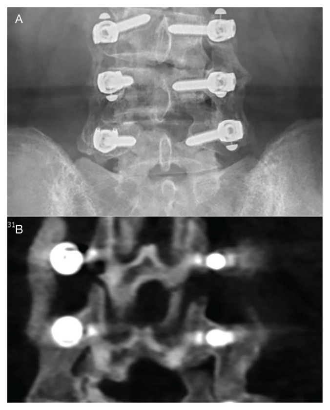
FIGURE 3.
Images Showing Arthrodesis
A, anteroposterior lumbar x-ray showing a solid arthrodesis bilaterally at L4-5 and a forming arthrodesis at the more recently operated L3-4 level. B, coronal computed tomography showing a solid arthrodesis bilaterally at L4-5 and a forming arthrodesis at the more recently operated L3-4 level.

