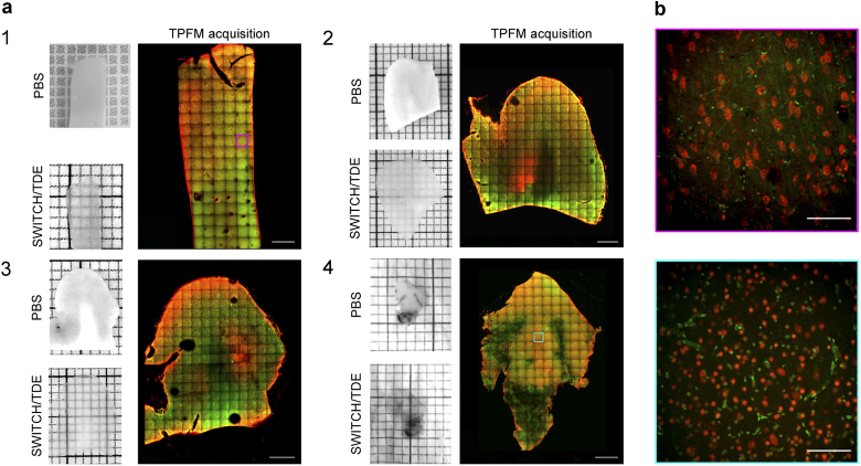
Fig. 2.
(a) Pictures showing the four analyzed human brain specimens before and after SWITCH/TDE clearing. A representative middle plane () of the mesoscopic reconstruction obtained with TPFM is shown next to each specimen. Scale bar = 1 mm. Specimens 1 and 2: two different portions of the left prefrontal cortex from adult and elderly subjects. Specimens 3 and 4: two surgically removed pieces from patients affected by Focal Cortical Dysplasia Type 2a (FCDIIa) and by Hemimegalencephaly (HME), respectively. (b) Magnified insets of specimen 1 (magenta) and 4 (cyan) showing the native resolution of the acquisition. Tissues were stained with an anti-NeuN antibody (in red) and with DAPI (in green). Scale bar =

