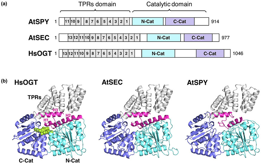
Figure 1. Structure comparison among human OGT, Arabidopsis SEC and SPY.
(a) Diagrams of HsOGT, AtSEC and AtSPY. TPRs are in grey. N-terminal catalytic domains, N-Cat, are in cyan. C-terminal catalytic domains, C-Cat, are in blue. (b) 3D structures of HsOGT (PDB ID: 4N3C, containing 4.5-TPRs)[71], and predicted 3D structures of Arabidopsis SEC and SPY using SWISS MODEL[72,73]. The HsOGT crystal structure (PDB ID: 4N3C)[71] was used as scaffold to predict AtSEC and AtSPY structures. The color schemes for HsOGT, AtSEC and AtSPY are as in (a). In (b), UDP-GlcNAc in HsOGT is shown as spheres (in lime-green). In the HsOGT structure in (b), the transitional helix (H3) between TPRs and N-Cat, and the first 2 α-helices (H1 and H2) of N-Cat are highlighted in magenta. The long intervening domain between N-Cat and C-Cat of HsOGT is omitted from the structure because this domain is uniquely present in the animal OGTs. This figure was modified from Zentella et al. [14].

