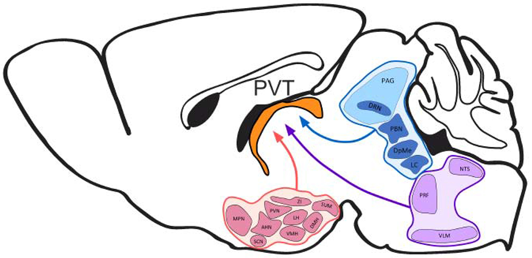
Figure 2. Schematic representation of major hypothalamic (pink), midbrain (blue), and hindbrain (purple) afferents to the rodent PVT (orange).

AHN, anterior hypothalamic nucleus; DMH, dorsomedial hypothalamus; DpMe, deep mesencephalic nucleus; DRN, dorsal raphe nucleus; LC, locus coeruleus; LH, lateral hypothalamus; MPN, medial preoptic nucleus; NTS, nucleus of the solitary tract; PAG, periaqueductal grey; PBN, parabrachial nucleus; PRF, pontine reticular formation; PVN, paraventricular hypothalamus; PVT, paraventricular thalamus; SCN, suprachiasmatic nucleus; SUM, supramammillary nucleus; VMH, ventromedial hypothalamus; VLM, ventrolateral medulla; ZI, zona incerta.
