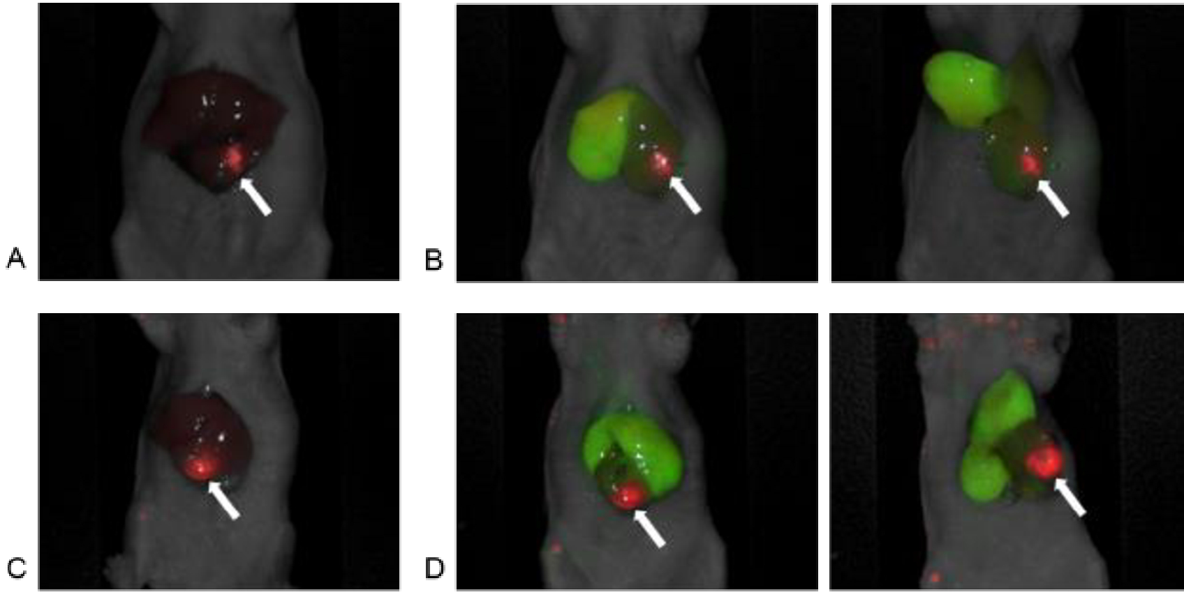
Figure 2.

In vivo imaging using the Pearl Trilogy Small Animal Imaging System. A) A representative image of red pseudo-color based on 700 nm signal of anti-CEACAM monoclonal antibody (6G5j) targeting surgically orthotopically implanted CM2 colon cancer liver metastatic patient tumor. Tumor-to-liver ratio (TLR) was 5.84. B) Red pseudo-color based on 700 nm signal of 6G5j targeting CM2 tumor and green pseudo-color based on 800 nm signal of ICG targeting the right lobe in the liver. Anatomic position and cranial inversion of the liver in the same mouse as Figure 2A were shown. C) Another representative image of red pseudo-color based on 700 nm signal of 6G5j targeting CM2 tumor. Tumor-to-liver ratio (TLR) was 3.88. D) Red pseudo-color based on 700 nm signal of 6G5j targeting CM2 tumor and green pseudo-color based on 800 nm signal of ICG targeting the right and the left medial lobe in the liver. Anatomic position and cranial inversion of the liver in the same mouse as Figure 2B were shown, (arrow: liver metastatic tumor)
