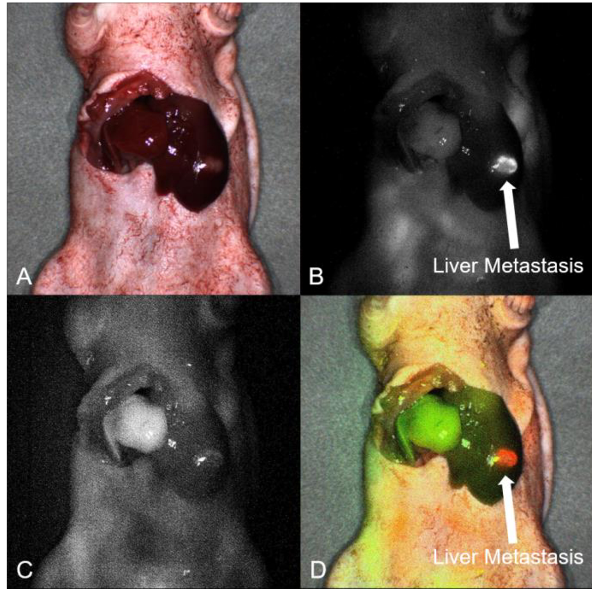
Figure 4.

Intra-vital dynamic imaging using the FLARE Imaging System. A) On the bright-light channel, the mice position was aligned. B) On the 700 nm channel, the surgically orthotopically implanted Liver5 colon cancer liver metastatic patient tumor had a clear fluorescence signal of anti-CEACAM monoclonal antibody (6G5j). (arrow: liver metastatic tumor). C) On the 800 nm channel, the left lobe had no fluorescence signal due to ligation of the left Glissonean pedicle and the right lobe in the liver had the clear fluorescence signal of ICG. D) Overlay mode showed clear differentiation between the tumor and segmental boundary on the same image, (arrow: liver metastatic tumor).
