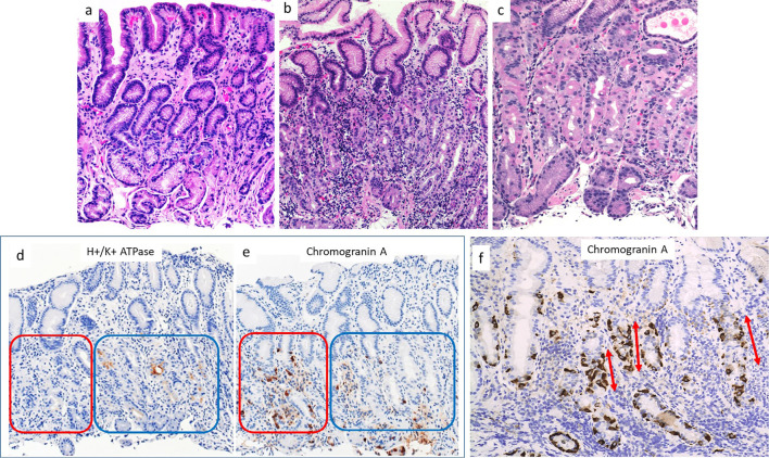
Figure 2.
Histopathological findings from various parts of the stomach in the patient in case 1. a) Tissue taken from the antrum. No inflammation or atrophy was present. b) Tissue taken from the greater curvature of the corpus. Lymphocytes and plasma cells were present in deeper, glandular tissue. c) Tissue taken from the lesser curvature of the corpus. Focal lymphocytic destruction of fundic glands was present in deeper tissue. Slight dilatation of oxyntic glands was observed, with parietal cell protrusion. d,e) Immunohistochemical staining for H+/K+ATPase and Chromogranin A. Parietal cells were lost on the left side (red square) but remained on the right side (blue square). ECL cells were more abundant in regions without parietal cells than in those with parietal cells (red square). f) Immunohistochemical staining for chromogranin A. Linear ECL cell hyperplasia was present (arrows).

