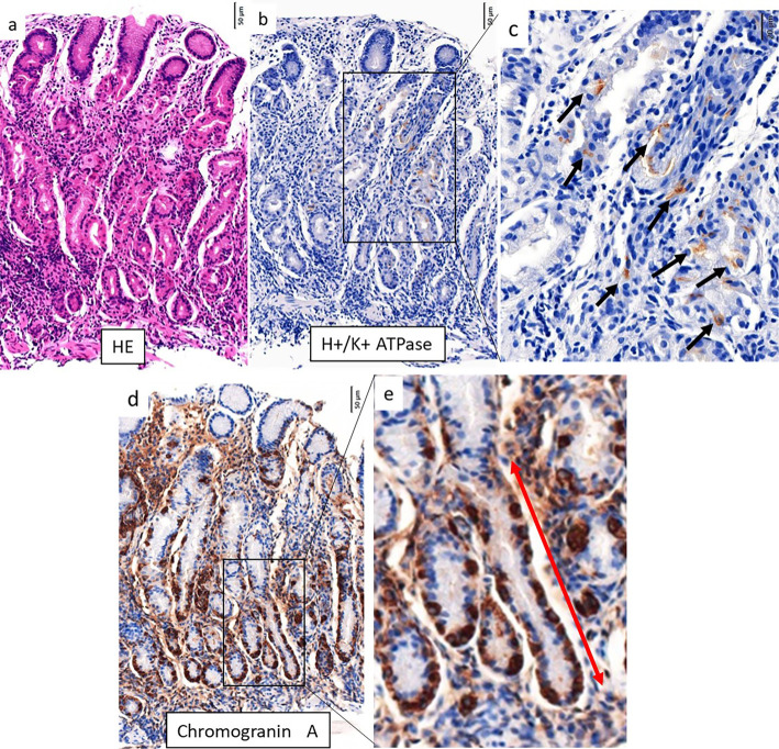
Figure 5.
Histopathological findings from the corpus in the patient in case 2. a) Lymphocytes and plasma cells were evident in deeper, glandular tissue. b, c) Immunohistochemical staining for H+/K+ATPase. There were only a few remaining parietal cells (black arrows). d, e) Immunohistochemical staining for Chromogranin A. Linear ECL cell hyperplasia was evident (red arrow).

