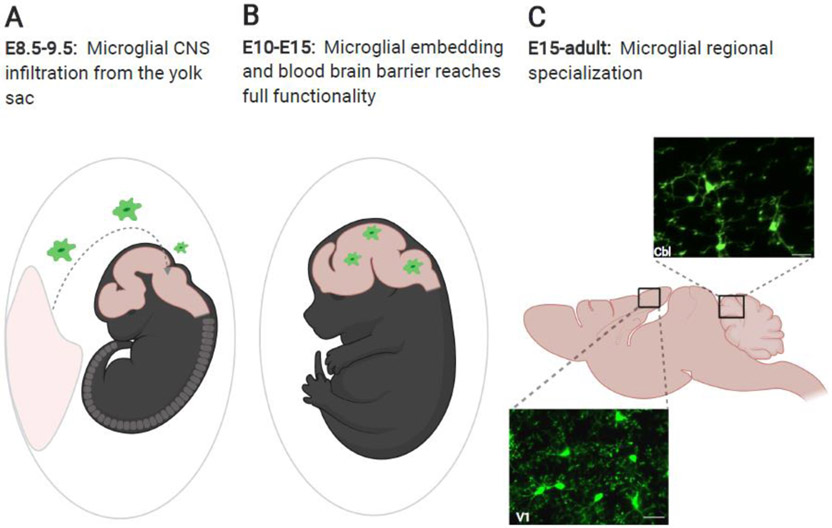
Figure 1: Microglial infiltration of the CNS and transformation by local cues.
(A) Microglia are born in the yolk sac and in mice, infiltrate the embryonic CNS between embryonic day (E)8.5 and E9.5 before the closure of the blood brain barrier. (B) Between E10 and E15 the blood brain barrier reaches full functionality, and microglia distribute within the CNS. (C) From E15 onward, influenced by local cues, microglia specialize to suit their local microenvironment. Pictured here are in vivo images of microglia from primary visual cortex (V1) and cerebellum (Cbl) in CX3CR1-GFP mice (unpublished images by Mark Stoessel and Ania Majewska). Scale bars = 25 μm. Figure created with Biorender.

