
Figure 7. Membrane encapsulation is essential for microvillus survival.
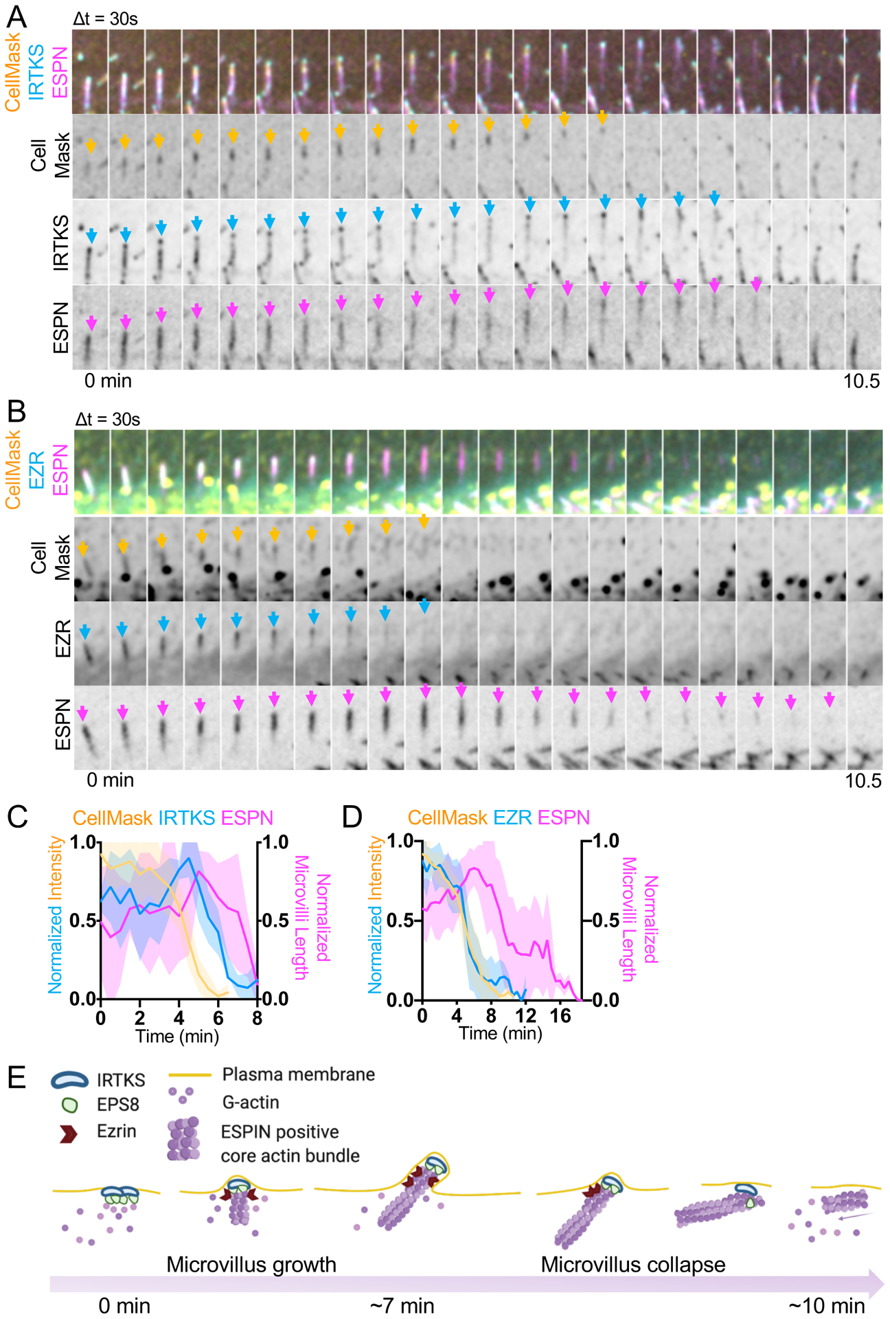
(A) Montage of a microvillus collapse event in a cell expressing EGFP-IRTKS and mCherry-ESPN, stained with CellMask-DeepRed. Box width = 2.5 μm. Related to Video S9. See also Figure S4D. (B) Montage of a microvillus collapse event in a cell expressing EZR-EGFP and mCherry-ESPN, stained with CellMask-DeepRed. Box width = 2.5 μm. Related to Video S10. See also Figure S4E. (C) Quantification of EGFP-IRTKS and CellMask-DeepRed intensity and microvilli length as represented in A; n = 4 events from 4 cells. (D) Quantification of EZR-EGFP and CellMask-DeepRed intensity and microvilli length vs. time. This plot was created by averaging traces of varying lengths ranging in duration from 6.5 – 12 min (EZR), 6 – 10.5 min (Cell Mask) and 7.5 – 18.5 min (ESPN); n = 15 events from 10 cells. For C and D, t = 0 is defined as −10 frames (5 min) before the drop in EGFP-IRTKS or EZR-EGFP intensity at the tips of microvilli. All images are represented as maximum intensity projections. For all curves, the solid line represents the mean and shading represents SD. (E) Model of de novo microvillus growth and collapse. Formation of EPS8 and IRTKS pucta coincide with local enrichment of G-actin followed by assembly of ESPN positive core actin bundles, recruitment of EZR, and plasma membrane protrusion. The process of de novo growth occurs on the scale of ~7 minutes. During microvillus collapse, nascent bundles lose membrane wrapping and enrichment of EZR in parallel, which in turn leads to loss of EPS8 and IRTKS, and subsequent collapse of the ESPN positive core actin bundle. The process of collapse also occurs on the scale of minutes. Model was created using BioRender. See also Figure S4.