Figure 1.
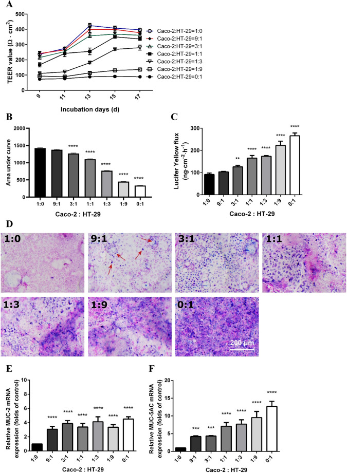
The monolayer integrity and mucus expression in the co-culture model at different ratios of Caco-2 and HT-29 cells. (A) TEER values of the cells grown on Transwell membranes. The cell cultures were subjected to TEER evaluation every other day since day 9. (B) Areas under the curves of TEER values of the co-culture monolayer calculated for statistical analysis. (C) Lucifer Yellow permeability assay of different combinations of Caco-2 and HT-29 cells at day 17. (D) Alcian Blue/nuclear fast red staining on Transwell membranes acquired by Nikon Plan 10 × /0.25 objective lens. Pink areas indicated nuclei and blue/purple areas indicated deposition of mucus. Mucin-related mRNA MUC-2 (E) and MUC-5AC (F) levels assessed by qRT-PCR. The target genes were normalized with reference gene β-actin. All values were presented as means ± SEM (N = 3, n = 3). Statistical differences were analyzed by Two-way analysis of variance (ANOVA), with Bonferroni post-hoc test. **p < 0.01, ***p < 0.001, ****p < 0.0001; significant different from Caco-2:HT-29 = 1:0 group.

