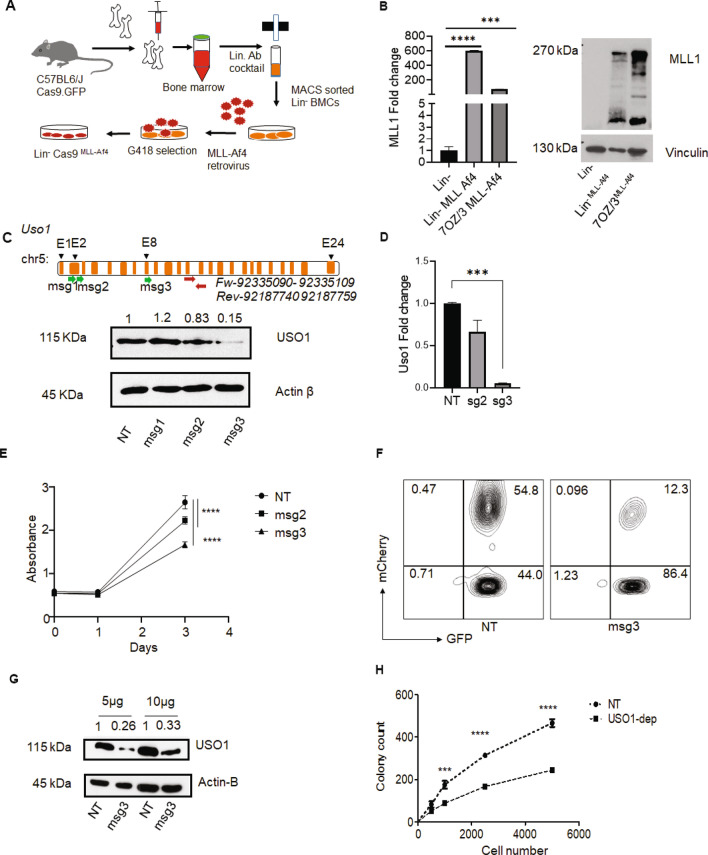
Figure 5.
USO1 depletion in transformed bone marrow cells shows reduced proliferation and colony forming potential. (A) Schematic of an in vitro model system to transform Lin- bone marrow cells from Cas9-egfp mice using overexpression of MLL-Af4 transgene in. (B) Analysis of overexpression of MLL-Af4 by RT-qPCR and western blot in retrovirally transduced Lin-Cas9MLL-Af4 cells, Lin-Cas9 cells were used as negative control and 70Z/3 cells transduced with MLL-Af4 were used as positive control. RT qPCR was performed with an optimized set of primers, normalized to L32, and represented as fold-change from an internal control for each experiment. Western Blotting was performed with an antibody to MLL1. Vinculin was used as a high molecular weight loading control. (C) Upper panel, schematic of murine Uso1 depletion experiments, showing location of sgRNAs relative to the gene, and RT-qPCR primer location. Bottom panel, western blot analysis of 70Z/3 cells transduced with three different sgRNAs targeting Uso1 (msg1-3) cloned in the MSCV.sgRNA.mCherry.v1 vector. (D) RT-qPCR analysis of Uso1 depletion in 70Z/3 cells. (E) MTS assay (Absorbance at 490 nm) to measure proliferation in Uso1-depleted 70Z/3 cells compared to NT control cells. (F) FACS plot showing gating schema to sort the Lin-Cas9MLL-Af4 GFP+ mCherry+ population following transduction with the Uso1 msg3 vector. (G) Western blot was used to confirm the reduction in USO1 expression in sorted Lin-Cas9MLL-Af4 cells. (H). Colony formation assay using Lin-Cas9MLL-Af4 cells in methylcellulose assay as described in methods with titration of input cell number (t test, ***P < 0.001; ****P < 0.0001).

