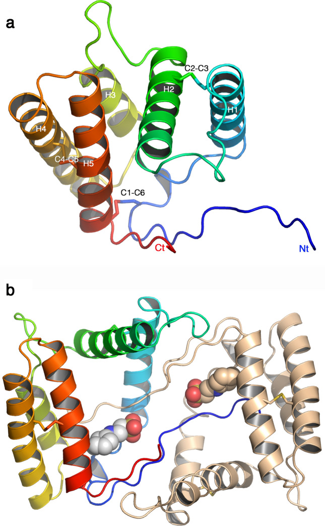
Figure 4.

Crystal structure of VdesOBP1 form P21. (a) Ribbon view of a monomer displaying helices 1–5 and disulphide bridges. The polypeptide chain is rainbow coloured, from blue (N-terminus) to red (C-terminus). The disulphide bonds are identified by the secondary structures they belong to. (b) Ribbon view of the crystallographic dimer. One monomer is rainbow coloured, the other one is coloured brown. The serendipitously bound buffer molecule 2-(N-cyclohexylamino)-ethane sulfonic acid (NCES) is displayed as atomic spheres (C: white, N: blue and O: red). Figure made with PyMOL (version 1.5.0.2. http://www.pymol.org).
