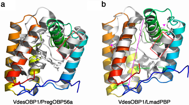
Figure 7.
Comparison of crystal structures of VdesOBP1 with the closest related OBPs from Phormia regina (PregOBP56) and Leucophera madereae (LmadPBP). (a) Ribbon view of the superposition of monomers from VdesOBP1 (rainbow coloured) and PregOBP56 (light grey) with its two fatty acid ligands (grey sticks). Note the absence of the classical OBP fold helix 2 (red squared) in VdesOBP1. (b) Ribbon view of the superposition of monomers from VdesOBP1 (rainbow coloured) and LmadPBP. Note the absence of the classical OBP fold helix 2 (red squared) in VdesOBP1 as compared to LmadPBP (red squared) and the shift of one disulphide bridge (red arrow). The 3D positions of the two other disulphide bridges are conserved (purple arrow), in the final fold, although as result of different C1-C6, C2-C3, C4-C5 (VdesOBP1) and C1-C3, C2-C5, C4-C6 (LmadPBP) connectivities. Figure made with PyMOL (version 1.5.0.2. http://www.pymol.org).

