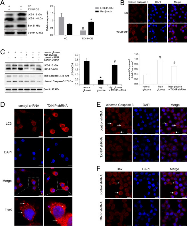
Fig. 3. TXNIP upregulation mediated high glucose-induced cell autophagy and apoptosis in RSC96 cells.
A Western blot and statistical analysis of the effect of TXNIP overexpression (OE) on LC3 and Bax in RSC96 cells. *P < 0.05 versus negative control group (NC). B Immunofluorescence of cleaved Caspase 3 in RSC96 cells transfected with negative control plasmid and TXNIP overexpression plasmid. Bar: 10 μm. C Western blot and statistical analysis of the effect of TXNIP knockdown using shRNA plasmid (pGenesil-1-TXNIP) on LC3 and cleaved Caspase 3 in RSC96 cells. *P < 0.05 versus normal glucose group. # means P < 0.05 versus high glucose group. D Immunofluorescence of LC3 in RSC96 cells transfected with control plasmid and TXNIP shRNA plasmid. The inset was shown with dashed square. White arrows showed the clustered LC3 expression. Bar: 10 μm. E Immunofluorescence of cleaved Caspase 3 in RSC96 cells transfected with control plasmid and TXNIP shRNA plasmid. White arrows showed the positive expression. Bar: 10 μm. F Immunofluorescence of Bax in RSC96 cells transfected with control plasmid and TXNIP shRNA plasmid. White arrows showed the positive signal. Bar: 10 μm.

