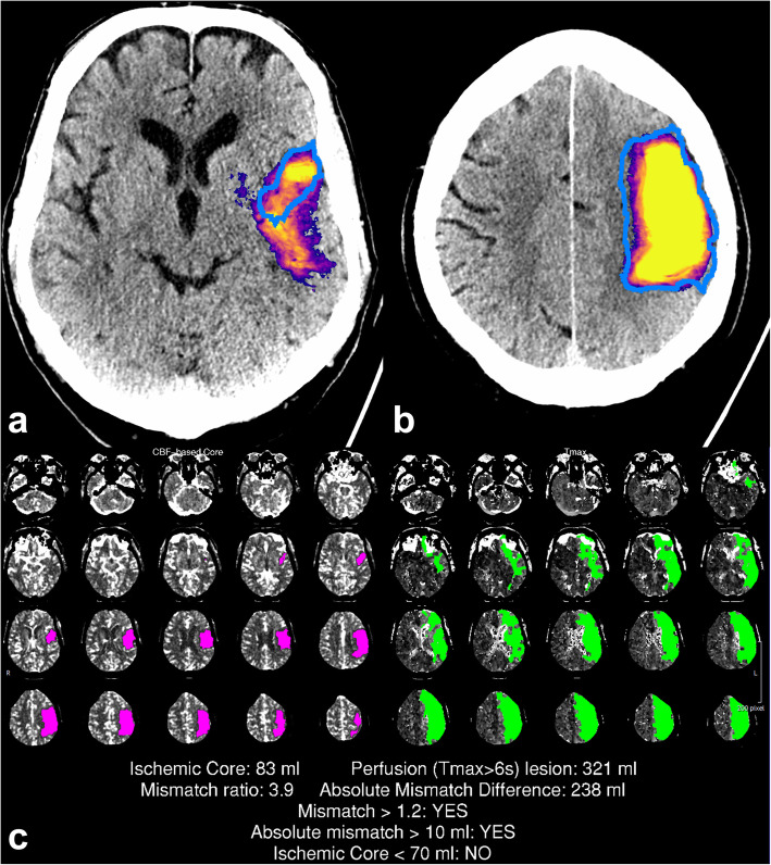
Fig. 2.
A large infarct correctly detected by the convolutional neural network (CNN). The CNN prediction included the final infarct (blue outline) and a part of the penumbra. Representative slices of the CNN predictions (a, b) with corresponding computed tomography perfusion RAPID (CTP-RAPID) report for comparison (c). The purple-orange-yellow colourmap depicts CNN output probability. Reported volumes: CTP-RAPID ischaemic core 83 mL, CNN 150 mL, and final infarct 150 mL

