
FIGURE 4.
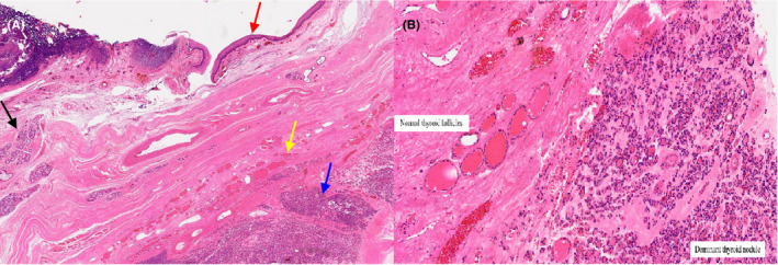
Microscopic histopathology of lingual thyroid specimen: A, Low magnification demonstrates oral type squamous mucosa (Red arrow), with subepithelial stroma, minor salivary glands (Black arrow), normal thyroid follicles (Yellow arrow), and a dominant thyroid nodule (Blue arrow); B, High magnification demonstrates thyroid tissue with features of nodule hyperplasia with a dominant (adenomatoid) partly calcified nodule within the subepithelial stroma and skeletal muscle