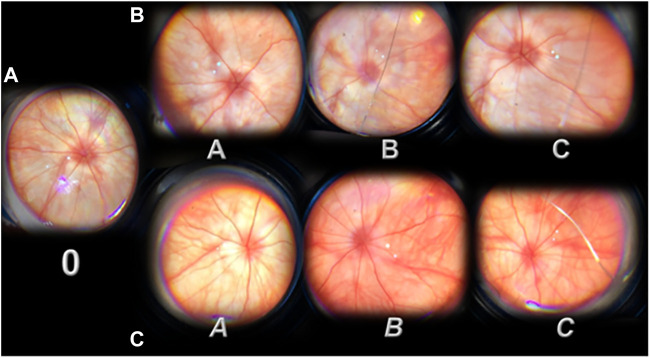
FIGURE 3.
Presentation of the rat retina after retrobulbar administered L-NAME (period from 20 minutes till the 1 day) before (0, (A)) and after therapy [saline (A, B, and C; (B)); BPC 157 (A, B, and C; (C))] administration. 0, L-NAME point before therapy application, at 20 min after retrobulbar administered L-NAME. Moderate generalized irregularity diameter blood vessels with moderate atrophy of the optic disk, faint presentation of the choroidal blood vessels (A). After therapy application (A, B, and C (saline) or A, B, and C (BPC 157)) (B,C). Immediately after (A, A). A. Strong generalized irregularity diameter blood vessels with severe atrophy of the optic disc, extremely poor presentation of the choroidal blood vessels (saline, B). A. Discrete generalized irregularity diameter blood vessels with mild atrophy of the optic disc, normal presentation of the choroidal blood vessels (BPC 157, C). 20 min after (B, B). B. Strong generalized irregularity diameter blood vessels with severe atrophy of the optic disc, extremely poor presentation of the choroidal blood vessels (saline, B). B. Normal eye background, normal presentation of the choroidal blood vessels (BPC 157, B,C). 1 day after (C,C). C. Strong generalized irregularity diameter blood vessels with severe atrophy of the optic disc, extremely poor presentation of the choroidal blood vessels (saline, B). C. Normal eye background, normal presentation of the choroidal blood vessels (BPC 157, C). The images are processed with software purchased with a USB microscope camera “Veho Discovery VMS-004 Deluxe.”

