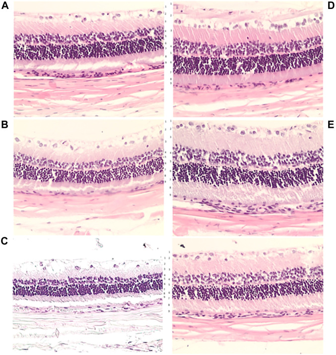
FIGURE 7.
Microscopic presentation of the rat retina at week 1 (A), week 2 (B) and week 4 (C) after L-NAME retrobulbar application, in controls (D) and BPC 157 treated rats (E), HE, x20. Transverse section of the retina (0.4–0.7 mm on the temporal side of the optic disc) showing a strict difference in the retinal layers and full retina thickness in the rats underwent L-NAME that received retrobulbar saline application and those that received BPC 157. More regular inner and outer nuclear layer and more regular distribution of ganglion cells, preserved thickness of the retina, the inner plexiform layer and inner nuclear layer at week 1 (A,E). More regular inner and outer nuclear layer and more regular distribution of ganglion cells, preserved thickness of the retina, the inner plexiform layer and inner nuclear layer at week 2 (B,E) (more regular inner and outer nuclear layer and more regular distribution of ganglion cells, preserved thickness of retina, the inner plexiform layer and inner nuclear layer). At week 4, BPC 157 treated rats show the preserved thickness of the whole retina, also inner plexiform layer and inner nuclear layer (C,E). Contrarily, degeneration of ganglion cells in control group is the most evident (C,D). Also, the outer nuclear layer is more regular in BPC 157 treated rats. 1—internal limiting membrane; 2—nerve fiber and ganglion cell layers; 3—inner plexiform layer; 4—inner nuclear layer; 5—outer plexiform layer; 6—outer nuclear layer; 7—outer limiting membrane; 8—photoreceptor layer; 9—pigment epithelium.

